Application Guide: Virus Characterization
Spectradyne’s ARC™ Particle Analyzer
Published:
Revision: V3
Overview
Spectradyne’s ARC™ particle analyzer is rapidly being adopted for virus quantification in research and GMP lab environments. It delivers fast viral titer measurements as well as single-particle molecular analysis using fluorescence for in-depth characterization.
The ARC enables accurate measurement of virus size and concentration, quantitative payload identification, and surface marker analysis directly in complex media without isolation or sample cleanup.
How the ARC Particle Analyzer Works
- Microfluidic Resistive Pulse Sensing (MRPS) directly measures single-particle size and concentration. MRPS is a non-optical electrical technique that is orthogonal to DLS, NTA, and flow cytometry.
- Single-particle fluorescence directly quantifies payload and surface ligands. Color multiplexing enables simultaneous measurements of encapsulated payloads, co-encapsulation, and targeting ligands.
Gold-Standard Accuracy for Size and Concentration
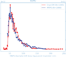
Direct comparison of MRPS and Cryo-TEM shows remarkable agreement across the full measurement range, validating the ARC’s accuracy for virus characterization.
Rapid Viral Titer in Complex Media
The ARC delivers simultaneous measurements of size, concentration, and fluorescence intensity. Up to three optical detection bands enable quantitative analysis of surface ligands and encapsulated payloads using standard fluorescence units.
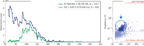
Quantifying RNA-Loaded Lentivirus
RNA-loaded lentivirus can be quantified directly using SYBR Gold fluorescence staining. Fluorescence-positive particles identify the loaded virion subpopulation within complex samples.
Compatibility with a Variety of Virus Samples
- Adenovirus
- Lentivirus
- Adeno-associated virus (AAV)
- SARS-CoV-2
- Influenza
- Herpes Simplex Virus (HSV)
- HIV, MLV, and other retroviruses
- Bacteriophage and viral aggregates