Applications
Extracellular Vesicles and Exosomes
Extracellular vesicles (EVs) are biological particles ranging in size from about 30 – 10,000 nanometers in diameter. Extracellular vesicles comprise diverse types of particles having a broad range of physical properties and biological origin, and include exosomes, ectosomes, microvesicles, microparticles, oncosomes, and apoptotic bodies. Extracellular vesicles have roles associated with an equally broad set of biological processes, including immune response, transfer of functional proteins and nucleic acid, elimination of unwanted materials, nutrition, surface-receptor mediated cell signaling, and numerous conditions of disease including cancer metastasis. Because of their involvement in so many physiological processes, extracellular vesicles hold enormous potential as therapeutics and biomarkers of health and disease. In these applications, accurate quantification of extracellular vesicles is critical for performing well controlled science, as well as ensuring safety and efficacy of extracellular vesicle-based products.
Accurate quantification with MRPS and simultaneous single-particle fluorescence
Spectradyne’s Arc delivers single-particle fluorescence combined with MRPS measurements, yielding size and phenotype for each individual nanoparticle, providing unparalleled individualized characterization not available with any other instrument. The Arc is fast and easy to use, and requires only 3 microliters of your sample to help you do better EV science.
See our recent Application Note on using the ARC to quantify immunostained extracellular vesicles.
Subpopulation analysis with fluorescence
Almost all biological nanoparticle samples contain heterogeneous populations of many different particle types. ARC can measure the size and concentration of the entire distribution using MRPS, and simultaneously measure and display only those particles in the sample that are positive for fluorescence in up to 3 different channels.

The example above shows that for an EV sample containing 1.26E+10 particles/ml overall, only 1.03E+09 particles/ml, or 8.1%, are tagged FITC- positive. In the same way, one can easily measure subpopulations in a virus sample, for instance.
Accurate quantification with microfluidic resistive pulse sensing (MRPS)
Spectradyne’s nCS1 delivers the most accurate concentration and size measurements of extracellular vesicles available in a practical bench top solution. The nCS1 is fast and easy to use, and requires only 3 microliters of your sample to help you do better EV science.
“The nCS1 is fast, accurate, and easy to use for routine EV quantification. We looked at other nanoparticle measurement solutions and the nCS1 is the only instrument that could measure the size of every nanoparticle of interest in as little as 3 uL of volume. Moreover, their customer service is superb and have went out of their way to ensure our nCS1 is always functioning well.”
-Joseph Sedlak, Co-Founder at Mercy BioAnalytics
Accurate concentrations yield better science
For measuring the biological activity of exosomes, characterizing vesicles of different biological origin and exploring vesicle biomarker applications, accurate concentration measurements are critical for performing well-controlled experiments.
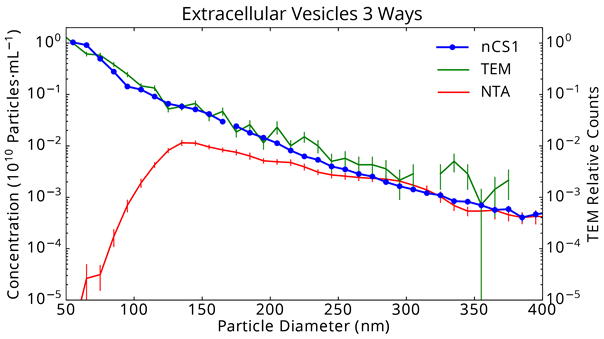
The figure below shows a comparison of Spectradyne’s nCS1 to NTA and the gold standard, Cryo-TEM. The nCS1 measurements are in excellent agreement with those of TEM, and indicate that the exosome concentration increases to smaller particle size down to 50 nm. In contrast, NTA reports misleading results: A loss of counting efficiency is apparent for particles as large as 200 nm in diameter, leading to a 10,000-fold error by 65 nm. Critically, NTA reports a prominent peak in the exosome size distribution near 130 nm diameter that does not in fact exist. Read more about why this happens.
More researchers are choosing Spectradyne’s instruments for extracellular vesicle quantification because these are fast and easy to use and deliver a more accurate analysis of their vesicles than they can obtain using any other method.
Contact us today for a free sample measurement or to request a demonstration!