Our ARCTM Particle Analyzer Delivers
Accurate Nanoparticle Size, Concentration, & Phenotype
Quantitative Single-Particle Fluorescence
Fast & easy to use, results in minutes
Watch and learn how the ARC can help your science
Real World Applications
Vesi-Ref CD63 GFP Analysis by Spectradyne’s ARC Particle Analyzer
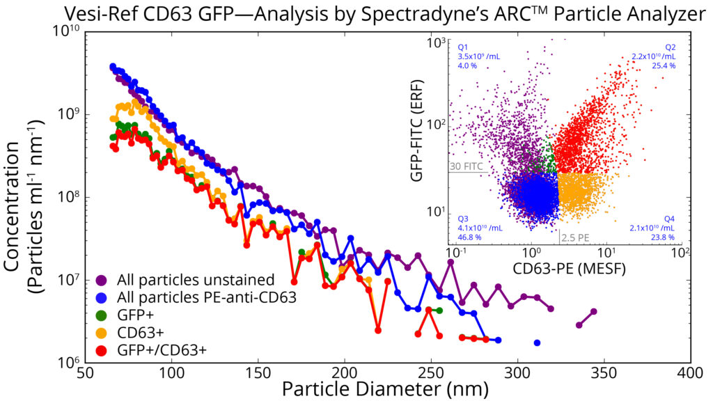
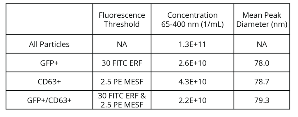
Fluorescence Microfluidic Resistive Pulse Sensing (F-MRPS, Spectradyne) was used to measure Vesi-Ref CD63 GFP (Vesiculab, UK) without modification and after staining with PE-anti-CD63. F-MRPS detects and measures the size and concentration of nanoparticles using MRPS, an electrical technique, while simultaneously measuring the fluorescence of each particle in up to 3 optical bands. Particle detection and fluorescent measurements are triggered using the MRPS signal, ensuring that each event represents a physical particle larger than the size limit of detection of the cartridge used (65 nm in this case). A one-time factory cross-calibration of the instrument to NIST-traceable MESF standard beads enables particle brightness to be reported by default in traceable units (ERF or MESF when appropriate).
The main figure shows particle concentration vs. size for all particles in the unstained sample (purple), all particles in the stained sample (blue), and subpopulations of the stained sample after gating GFP+ (green), CD63+ (yellow), and double-positive GFP+/CD63+ (red) populations. While the overall particle size distribution displays an approximate power-law dependence of concentration on size that extends to the lower size limit of detection for this cartridge, the fluorescent subpopulations each show the presence of a peak near 75 nm diameter. The 2D scatter plot (inset) shows the brightness of each particle in FITC and PE channels. Absolute concentration and relative abundance of particles are reported for each quadrant, the bounds of which are defined by the fluorescence limits of detection of each channel: 30 FITC ERF and 2.5 PE MESF.
Learn more at Vesiculab.com!
Check out this video to learn more
The Arc particle analyzer simultaneously measures single-particle fluorescence in up to three optical channels while leveraging the proven strengths of microfluidic resistive pulse sensing (MRPS) to quantify size and absolute concentration. The result: Fast and accurate quantification of target populations in the most challenging biological samples.
Quantify Fluorescent Sub-populations with ease
The GFP recombinant extracellular vesicles were mixed with a significantly higher concentration of non-fluorescent extracellular vesicles isolated from tissue culture. While the overall particle size distribution is dominated by that of the non-fluorescent background EVs, the fluorescent GFP-rEVs are clearly identified in the mixture by their fluorescence.
