Extracellular Vesicles and Exosomes
Size, Concentration, and Cargo: Accurate measurement for well-controlled science
Accurate particle size and concentration
Cargo analysis by single-particle fluorescence
Fast and easy to use: No alignment, calibration, or cleaning required

See how our F-MRPS technology stacks up against other methods in Extracellular Vesicle (EV) applications

Accurate EV quantification: Size, Concentration, Targeting, and Cargo
Extracellular vesicles (EVs) are biological nanoparticles that hold enormous promise for their potential as therapeutics and as biomarkers of health and disease. At this size scale, the size and concentration of EVs directly impact their behavior: Size determines cargo capacity and diffusion rate, (e.g., bioactivity) and concentration represents the total amount of cargo in an EV preparation (i.e., the dose, in therapeutic terms). Controlled therapeutic delivery via surface markers are vital to ensure proper targeting and regulated immunogenicity. Particle size, concentration, targeting, and cargo therefore represent four critical quality attributes of EVs that must be accurately measured to understand and develop the potential of EVs.
Extracellular vesicles are produced by cells in complex, heterogeneous environment, and the isolation of EVs from these complex mixtures remains a time consuming, challenging, and still imperfect science in itself. Accurate EV quantification therefore also requires the ability to distinguish the particles of interest i.e., phenotype, from a complex, heterogeneous background.
Common light scattering-based optical techniques such as dynamic light scattering (DLS), nanoparticle tracking analysis (NTA) and nanoscale flow cytometry (nanoFCM) are insufficient for this task. These techniques require assumptions of monodisperse size or homogeneous optical properties throughout the sample (i.e., uniform particle refractive index) that cannot be justified for these preparations.
Accurate and True: EV Size & Concentration with MRPS

The figure above demonstrates the accuracy of MRPS for EV size and concentration measurement by comparing to the gold standard for particle size measurement, transmission electron microscopy (TEM). The power law dependence of concentration on size measured by TEM is identically reproduced by the MRPS method while nanoparticle tracking analysis (NTA) falls far short.
Why don’t the NTA measurements agree with TEM and MRPS? NTA and other light scattering-based techniques lose sensitivity to small particles when larger ones are present in the sample. If that sounds like a surprise, convince yourself with a simple experiment you can do on your own NTA.
Look beyond light scattering
Why assume anything at all about your particles when you don’t have to? Microfluidic resistive pulse sensing (MRPS) delivers accurate measurements of nanoparticle size, concentration, and absolute fluorescence without any a priori assumptions about the sample or particle composition. Because MRPS measures particles one-by-one, independently of one another, it accurately measures particles of any polydispersity. Because MRPS directly measures each particle’s size (its volume to be precise)—electrically—it accurately measures particles of any material. And because Spectradyne’s ARC particle analyzer does all the optical alignment for you, continuously throughout measurement, it delivers repeatable, and quantitative fluorescence measurements without any calibration.
When the experimental control of your science is on the line, you need more than light scattering alone.
Quantitative subpopulation analysis
Almost all biological nanoparticle samples contain heterogeneous populations of many different particle types. Spectradyne’s ARC particle analyzer measures the size and concentration of the entire distribution using MRPS, and simultaneously measures and displays only those particles in the sample that are positive for fluorescence in up to 3 different channels.

The example above demonstrates the quantification of a subpopulation of fluorescent extracellular vesicles outnumbered by a broad background of non-fluorescent EVs. Results are quantitative over any size range: On the range of 80-150 nm diameter, fluorescent EVs are present at 1×10⁹ particles/mL and represent 8% of all particles in that same size range (1.3×10¹⁰ particles/ml).
EV Phenotyping – Truly simultaneous multicolor fluorescence

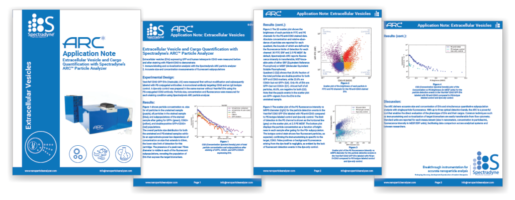
Spectradyne’s ARC particle analyzer was used to characterize synthetic extracellular vesicles expressing GFP and the human tetraspanin CD63 (Vesi-Ref CD63 GFP, Vesiculab, UK). The ARC delivers accurate size, concentration, and quantitative fluorescence in absolute units for each particle and enables the complete and thorough characterization of the sample in a few minutes. Read the full application note here.
How our MRPS technology stacks up against DLS and NTA

Spectradyne’s nCS1TM and ARCTM deliver the most accurate concentration and size measurements of extracellular vesicles available in a practical bench top solution, combined in the ARC with fluorescence measurements. Spectradyne’s nanoparticle analyzers are fast and easy to use, and require only 3 microliters of your sample to help you do better EV science.