
One of the major benefits of Microfluidic Resistive Pule Sensing (MRPS) is that MRPS directly measures particle volume. This sizing method is quite different from light-scattering based techniques, which first assume that all particles are spherical in shape, and then infer particle diameter from some other physical property being measured (e.g., mean squared displacement of diffusion).
Back to table of contents
In MRPS, the measured electrical response is directly proportional to the volume of the particle occluding the nanoconstriction. In order to easily display particle size results, however, the volume of each particle is converted to an Equivalent Spherical Diameter (ESD). Particle concentration is then plotted against ESD, as is typical for all particle analysis systems. The important thing to remember is that in RPS, the particle size (volume) is measured directly and stored, whereas in light scattering systems particle size is calculated from diffusion behavior assuming particles are spherical.
To show how this characteristic of MRPS can manifest, the following example may be useful. A customer-supplied virus sample was measured in the nCS1 MRPS system. A clear peak in the distribution is seen for the monomer at 136 nm (see figure to right). A small secondary "hump" can also be seen at around ~170 nm, as shown to the right in the volume-weighted particle size distribution.
The hump in the data seen at 170 nm indicates the presence of virus dimers. Remembering that the nCS1 measures volume, and that ESD scales as the cube root of volume, a dimer will have twice the volume of the monomer, corresponding to ~1.25 times the ESD of the monomer. In this case, with a monomer at 136 nm ESD, the dimer appears right where it is expected, at 170 nm.
Another interesting example is provided by a customer-supplied virus that was cylindrical in shape. The customer had TEM data showing the virus to be a cylinder of diameter ~65 nm and length of ~180 nm. Given these dimensions, we calculate the volume of the cylinder, and then the ESD of a sphere of same volume, which predicts an ESD of approximately 100 nm for the virus. As seen in the second graph to the right, this is exactly what the nCS1 measures!
The nCS1 MRPS system offers higher resolution to begin with, but the fact that it measures volume directly also yields more valuable insight into particle size distributions as well!
Please continue to follow our blog as we share insights, technical details, and generally geek-out with you about nanoparticle science!
Email us for more information, or to discuss your particular application directly.
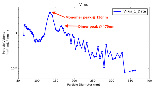
A virus sample measured with Spectradyne's nCS1TM, showing monomer and dimer virus particles.
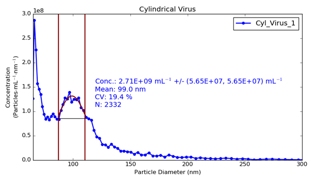
Cylindrical virus particles plotted in terms of ESD.

Spectradyne is a world leader in the microfluidic measurement of nanoparticles.
Nanoparticle analysis using resistive pulse sensing avoids many of the difficulties associated with DLS and optical tracking. Spectradyne is the only company providing microfluidic particle size analysis, now combined with single particle fluorescence.
Check us out today!