
Learn from NIH and Johns Hopkins scientists as they share the importance of accurate extracellular vesicle quantification in their research:
Our library is expanding! Watch an updated introduction to our technology, see a virtual demo, take a dive into our microfluidic cartridges, or learn different ways to measure concentration using Spectradyne's nCS1.
Stay tuned for more additions in the near future!
Spectradyne is a proud sponsor of the 2020 American Association of Pharmaceutical Scientists PharmSci 360, October 26-November 5, 2020. We will host a virtual exhibit booth and present a poster describing the advantages of using MRPS to quantify virus entitled "Non-Biological Quantification of Viral Titer: A New Approach." If you can't attend, you will be able to view the poster in our library after the show.

Ever wondered what's in the sunscreen you used all summer? Which brand provides the best value on a dollars-per-particle basis? We used Spectradyne's nCS1 to find out!
Read about the experiments and see the results in our blog post
How do researchers use Microfluidic Resistive Pulse Sensing (MRPSTM) to make scientific discoveries?
Here are some examples:
Scientists at John Hopkins University and collaborators at the National Institute of Standards and Technology (NIST) recently released a pre-print article comparing emerging tools for quantifying Extracellular Vesicles (EVs). The study compared Nanoparticle Tracking Analysis (NTA), Single-Particle Interferometric Reflectance Imaging Sensing (SP-IRIS), nano Flow Cytometry and Spectradyne's Microfluidic Resistive Pulse Sensing (MRPS).
E. Mallick et al., "Characterization of extracellular vesicles and artificial nanoparticles with four orthogonal single-particle analysis platforms" doi: 10.1101/2020.08.04.237156
Massachusetts Institute of Technology scientists describe the investigation and measurement of chiral supraparticles with applications to drug delivery systems, tumor detection markers, biosensors, and other biomaterial-based devices.
Yeom, P. P. G. Guimaraes, H. M. Ahn, B.-K. Jung, Q. Hu, K. McHugh, M. J. Mitchell, C.-O. Yun, R. Langer, A. Jaklenec, "Chiral Supraparticles for Controllable Nanomedicine," Advanced Materials 32, 1903878 (2019) doi: 10.1002/adma.201903878
Researchers at Aston University in Birmingham, UK, used MRPS to characterize lipid nanoparticles:
A. J. Rozoa, M. H. Coxa, A. Devitta, A. J. Rothniea, A. D. Goddard, "Biophysical analysis of lipidic nanoparticles," Methods doi.org/10.1016/j.ymeth.2020.05.001
A paper from Zoltan Varga's group at the Research Center for Natural Sciences (Budapest, Hungary) describes complementary methods for measuring silica nanoparticles.
M. A. Al-Khafaji, A. Gaal, A. Wacha , A. Bota, Z. Varga, "Particle Size Distribution of Bimodal Silica Nanoparticles: A Comparison of Different Measurement Techniques," Materials 13, 3101 (2020) doi.org/10.3390/ma13143101
A paper from the lab of Dr. Yvonne Perrie at University of Strathclyde describes using MRPS to characterize novel liposomes for activated drug delivery.
C. B. Roces, E. C. Port, N. N. Daskalakis, J. A. Watts, J. W. Aylott, G. W. Halbert, Y. Perrie, "Rapid scale-up and production of active-loaded PEGylated liposomes," Intl. J. Pharmaceutics 586, 119586 (2020) doi.org/10.1016/j.ijpharm.2020.119566
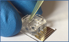
Many of you have never seen what it looks like to operate the nCS1, so we put together a short video demonstration. This 13-minute video will walk you through a "virtual demo" of the nCS1: from loading the sample into a microfluidic cartridge to producing a final report. You will see how easy it is to get high quality, high resolution nanoparticle size and concentration data from the nCS1 in just minutes. Once you've seen this video, you will be ready to send us samples to see your own data in high definition!

Spectradyne is a world leader in the microfluidic measurement of nanoparticles.
Nanoparticle analysis using resistive pulse sensing avoids many of the difficulties associated with DLS and optical tracking. Spectradyne is the only company providing microfluidic particle size analysis, now combined with single particle fluorescence.
Check us out today!